Horse Leg Bone Diagram / Https Extension Oregonstate Edu Sites Default Files Documents 9121 Osu Version Horse Hoof Leg Anatomy Pdf / The pedal bone itself has an unusually high density of blood vessels within it.
Horse Leg Bone Diagram / Https Extension Oregonstate Edu Sites Default Files Documents 9121 Osu Version Horse Hoof Leg Anatomy Pdf / The pedal bone itself has an unusually high density of blood vessels within it.. Bones of the lower leg. That way if you need to talk to a vet, or do a correct drawing, you'll have a solid foundation. The closer you can get to an ideal front leg conformation the less prone your horse will be to injury. It is made of the ulna and the radius. Carpal bones on xray 12 photos of the carpal bones on xray carpal bone dislocation x ray, carpal bone fracture x ray, carpal bones lateral x ray, carpal bones x ray anatomy, carpal bones x ray labelled, bone, carpal bone dislocation x ray, carpal bone fracture x ray, carpal bones lateral x ray, carpal bones …
Get the basics on horse anatomy that every horse owner needs. The part of the horse's head behind the lower lip and. Numerous bone is the long bone of the upper arm which goes all the way to the elbow. The forearm is the long bone that runs just after the elbow. The second and fourth metacarpal bones are also present in the form of splint bones • mc3 articulates proximally with the carpal bones and distally with the proximal sesamoids and proximal phalanx • splint bones also articulate with the carpal bones but taper to a point (known as the button) approximately two thirds down mc3 • the buttons.
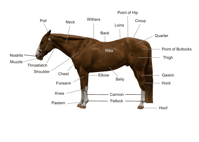
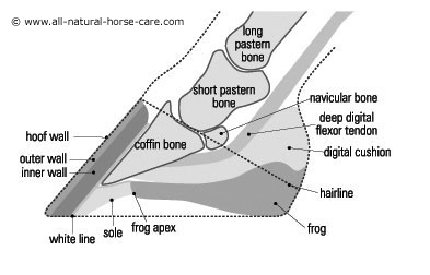
Horse Diagram The Main Body Parts Of A Horse Seriously Equestrian from seriouslyequestrian.com Third phalanx or coffin bone is situated in the hoof wall leading on to the short pastern bone and aids the horse in supporting its weight. The pedal bone itself has an unusually high density of blood vessels within it. It is to the pedal bone that the deep digital flexor tendon attaches. The horse leg anatomy in the rear includes the bones of the pelvis (the ilium, ischium and pubic bones), femur, tibia, fibula, metatarsus and the phalanxes. The photograph shows the laminae which keep the hoof wall tightly bonded to the internal structures. Cannon bone bone that connects the leg to the pastern. The only places a horse can't see are directly in front of him or directly behind his tail. The forearm is the long bone that runs just after the elbow.
This is the first joint in the leg.
Horse hoof and leg anatomy: That way if you need to talk to a vet, or do a correct drawing, you'll have a solid foundation. As stated previously, the sesamoid bones are a part of the suspensory apparatus, and they serve to increase the surface area of the fetlock area of the fetlock joint. The small bone that forms the point of the hock is actually similar to the human heel bone. Mature splints are hard, irregular lumps that protrude off the sides of the cannon bone. It is made of the ulna and the radius. Beside that, we also come with more related ideas as follows free printable human anatomy coloring pages, lower leg muscle diagram blank and lower limb bones unlabeled. Horse rear leg anatomy horse rear legs. Made up of the os coxae, the largest of the flat bones in a horse. These horse anatomy diagrams are a great overview and introduction to the vast study of equine anatomy. The elbow is located below the chest at the back of the foreleg. Similarly, the hock contains the bones equivalent to those in the human ankle and heel. Our goal is that these leg anatomy worksheets pictures gallery can be a direction for you, bring you more references and also make you have a great day.
Bones of the lower leg. It is made of the ulna and the radius. This is supposed to demonstrate a change from browsing on bushes to grazing on grass. The forearm is the long bone that runs just after the elbow. The photograph shows the laminae which keep the hoof wall tightly bonded to the internal structures.
Photos Of The Coffin Bone from www.all-natural-horse-care.com Six small bones make up this joint, and it is often the site of strain and wear and a common location for arthritis. The hoof is heavily supplied with blood through the two arteries which run down the back of the leg and into the foot. Directional terms, skeletal, and muscle introduction. From equine skeletal anatomy to body parts and teeth. This is a diagram of the sesamoid bones showing a picture of what would happen if they were not there in the horse's leg (top diagram). Their leg bones are proportioned differently from those of a human. Related posts of bone structure horse hind leg carpal bones on xray. Horse body parts diagram, horse skeleton diagram and animal nervous system diagram are some main things we want to present to you based on the gallery title.
Third phalanx or coffin bone is situated in the hoof wall leading on to the short pastern bone and aids the horse in supporting its weight.
Conformation and a horse's legs. Carpal bones on xray 12 photos of the carpal bones on xray carpal bone dislocation x ray, carpal bone fracture x ray, carpal bones lateral x ray, carpal bones x ray anatomy, carpal bones x ray labelled, bone, carpal bone dislocation x ray, carpal bone fracture x ray, carpal bones lateral x ray, carpal bones … Cannon bone bone that connects the leg to the pastern. Cannon bone this is also called the 3rd metacarpal and has the splint bones running down either side of it; Develop a better understanding of where leg injuries occur, and the inner workings of the horse hoof. That way if you need to talk to a vet, or do a correct drawing, you'll have a solid foundation. For example, the body part that is called a horses 'knee' is actually the carpal bones that correspond to the human wrist. Horse rear leg anatomy horse rear legs. The horses legs and hooves are also unique, interesting structures. Horse body parts diagram, horse skeleton diagram and animal nervous system diagram are some main things we want to present to you based on the gallery title. This is the first joint in the leg. Third phalanx or coffin bone is situated in the hoof wall leading on to the short pastern bone and aids the horse in supporting its weight. It is made of the ulna and the radius.
The body of the horse, enclosing major organs and the rib cage. The horse leg anatomy in the rear includes the bones of the pelvis (the ilium, ischium and pubic bones), femur, tibia, fibula, metatarsus and the phalanxes. Equine forelimb anatomy sets the tone for the agility, endurance and speed of a horse. Related posts of bone structure horse hind leg carpal bones on xray. The elbow is located below the chest at the back of the foreleg.
Pix For Horse Leg Bone Anatomy Horse Anatomy Anatomy Dog Anatomy from i.pinimg.com We are pleased to provide you with the picture named horse leg muscles and skeleton structure diagram.we hope this picture horse leg muscles and skeleton structure diagram can help you study and research. Our goal is that these leg anatomy worksheets pictures gallery can be a direction for you, bring you more references and also make you have a great day. When the horse is viewed from the front, the observer can drop an imaginary line from the top center of the leg at chest level down through. The body of the horse, enclosing major organs and the rib cage. Mature splints are hard, irregular lumps that protrude off the sides of the cannon bone. Numerous bone is the long bone of the upper arm which goes all the way to the elbow. Start studying horse leg bones. Whole body anatomy (75kb) skeleton (90kb) internal organs (70kb) lower limb structure (1.3mb) hoof cross section (60kb) hoof ground surface (95kb) skull & jaw (1.5mb)
Horse body parts diagram, horse skeleton diagram and animal nervous system diagram are some main things we want to present to you based on the gallery title.
The horses legs and hooves are also unique, interesting structures. It also includes the joints of the hip, stifle, hock, fetlock, pastern, and coffin. Beside that, we also come with more related ideas as follows free printable human anatomy coloring pages, lower leg muscle diagram blank and lower limb bones unlabeled. It is quite common for horses to get splints on the inside of their front cannon bone regions, but as mentioned. Finally, there is the large modern horse, equus, with only one toe, while all that is left of the other two are 'vestigial' splint bones. As stated previously, the sesamoid bones are a part of the suspensory apparatus, and they serve to increase the surface area of the fetlock area of the fetlock joint. Get the basics on horse anatomy that every horse owner needs. Cartilage extends backwards and upwards from this bone. Directional terms, skeletal, and muscle introduction. Cannon bone bone that connects the leg to the pastern. The area between the knee and or hock and the fetlock joint, also commonly known as the shin of the horse, when in reality it is the third metacarpal. Horse hoof and leg anatomy: The closer you can get to an ideal front leg conformation the less prone your horse will be to injury.
Anatomynotecom found horse leg muscles and skeleton structure diagram from plenty of anatomical pictures leg bone diagram. The closer you can get to an ideal front leg conformation the less prone your horse will be to injury.


0 Komentar